 Urinary or Urethral catheterization is a process in which a urinary catheter (such as a Foley catheter) is either inserted through a female patient's urinary tract into their bladder or attached to a male patient's penis. A balloon located at the end of the catheter is usually inflated with sterile water to prevent the catheter from slipping out. In this manner, the patient's urine is collected and contained for various medical purposes. The procedure of catheterization will usually be done by a clinician, often a nurse, although self-catheterization is possible as well. Urinary catheterization is a routine medical procedure that has both diagnostic and therapeutic purposes. Types of CatheterizationCatheters come in a large variety of sizes; materials (latex, silicone, PVC, or Teflon); and types (Foley catheter, straight catheter, or coude tip catheter). In the case of internal catheters, those inserted into the urethra, the smallest size is usually recommended, although a larger size is sometimes needed to control leakage of urine around the catheter. A large size can also become necessary when the urine is thick, bloody or contains large amounts of sediment. Larger internal catheters, however, are more likely to cause damage to the urethra. Some people have developed allergies or sensitivities to latex after long-term latex catheter use. In such cases, silicone or Teflon types should be used. Proper catheter use can also often be determined by the length of time for which the process is necessary: long-term (often called indwelling) or short-term use. Sex DifferencesIn males, the catheter tube is inserted into the urinary tract through the penis. A condom catheter can also be used. In females, the catheter is inserted into the urethral meatus, after a cleansing using povidone-iodine. The procedure can be complicated in females due to varying layouts of the genitalia (due to age, obesity, Female genital cutting, childbirth, or other factors), but a good clinician should rely on anatomical landmarks and patience when dealing with such a patient. IndicationDiagnostic o Collection of uncontaminated urine specimen o Monitoring of urine output o Imaging of the urinary tract Therapeutic o Acute urinary retention (eg, benign prostatic hypertrophy, blood clots) o Chronic obstruction that causes hydronephrosis o Initiation of continuous bladder irrigation o Intermittent decompression for neurogenic bladder o Hygienic care of bedridden patients ContraindicationUrinary catheterization is contraindicated in the presence of traumatic injury to the lower urinary tract (eg, urethral tear). This condition may be suspected in male patients with a pelvic or straddle-type injury. Signs that increase suspicion for injury are a high-riding or boggy prostate, perineal hematoma, or blood at the meatus. When any of these findings are present in the setting of concerning trauma, a retrograde urethrogram should be performed to rule out a urethral tear prior to placing a catheter into the bladder. AnesthesiaTopical anesthesia is administered with lidocaine gel 2%. Many facilities have a preloaded syringe with an opening appropriate for insertion into the meatus available either separately or in the catheter kit. To instill, hold the penis firmly and extended, place the tip of the syringe in the meatus, and apply gentle but continuous pressure on the plunger. EquipmentCommercial single-use urethral catheterization tray o Povidone iodine o Sterile cotton balls o Water-soluble lubrication gel o Sterile drapes o Sterile gloves o Urethral catheter o Prefilled 10-mL saline syringe o Urinometer connected to a collection bag Sterile anesthetic lubricant (eg, lidocaine gel 2%) with a blunt tip urethral applicator or a plastic syringe (5-10 mL) PositioningPlace the patient supine, in the frogleg position, with knees flexed. Technique· Explain the procedure, benefits, risks, complications, and alternatives to the patient or the patient's representative. · Position the patient supine, in bed, and uncover the genitalia. · Open the catheter tray and place it on the gurney in between the patient's legs; use the sterile package as an extended sterile field. Open the iodine/chlorhexidine preparatory solution and pour it onto the sterile cotton balls. Open a sterile lidocaine 2% lubricant with applicator or a 10-mL syringe and sterile 2% lidocaine gel and place them on the sterile field. · Wear sterile gloves and use the nondominant hand to hold the penis and retract the foreskin (if present). This hand is the nonsterile hand and holds the penis throughout the procedure. · Use the sterile hand and sterile forceps to prep the urethra and glans in circular motions with at least 3 different cotton balls. Use the sterile drapes that are provided with the catheter tray to create a sterile field around the penis. · Using a syringe with no needle, instill 5-10 mL of lidocaine gel 2% into the urethra. Place a finger on the meatus to help prevent spillage of the anesthetic lubricant. Allow 2-3 minutes before proceeding with the urethral catheterization. · Hold the catheter with the sterile hand or leave it in the sterile field to remove the cover. Apply a generous amount of the nonanesthetic lubricant that is provided with the catheter tray to the catheter. · While holding the penis at approximately 90º to the gurney and stretching it upward to straighten out the penile urethra, slowly and gently introduce the catheter into the urethra. Continue to advance the catheter until the proximal Y-shaped ports are at the meatus. · Wait for urine to drain from the larger port to ensure that the distal end of the catheter is in the urethra. The lubricant jelly–filled distal catheter openings may delay urine return. If no spontaneous return of urine occurs, try attaching a 60-mL syringe to aspirate urine. If urine return is still not visible, withdraw the catheter and reattempt the procedure (preferably after using ultrasonography to verify the presence of urine in the bladder). · After visualization of urine return (and while the proximal ports are at the level of the meatus), inflate the distal balloon by injecting 5-10 mL of 0.9% NaCl (normal saline) through the cuff inflation port. Inflation of the balloon inside the urethra results in severe pain, gross hematuria, and, possibly, urethral tear. · Gently withdraw the catheter from the urethra until resistance is met. Secure the catheter to the patient's thigh with a wide tape. Creating a gutter to elevate the catheter from the thigh may increase the patient's comfort. If the patient is uncircumcised, make sure to reduce the foreskin, as failure to do so can cause paraphimosis. PearlsInsertion of a Coudé catheter: The Coudé catheter, which has a stiffer and pointed tip, was designed to overcome urethral obstruction that a more flexible catheter cannot negotiate (eg, patients with benign prostatic hypertrophy). To place a Coudé catheter, follow the procedure described above. The elbow on the tip of the catheter should face anteriorly to allow the small rounded ball on the tip of the catheter to negotiate the urogenital diaphragm. Perineal pressure assistance: The distal tip of the catheter might become caught in the posterior fold between the urethra and the urogenital diaphragm. An assistant can apply upward pressure to the perineum while the catheter is advanced to direct the catheter tip upward through the urogenital diaphragm. Complications-Infections o Urethritis o Cystitis o Pyelonephritis o Transient bacteremia -Paraphimosis, caused by failure to reduce the foreskin after catheterization -Creation of false passages -Urethral strictures -Urethral perforation -Bleeding Open Section* Prophylactic antibiotics are recommended for patients with prosthetic heart valves, artificial urethral sphincters, or penile implants. * Catheter types and sizes o Adults: Foley (16-18 F) o Adults with obstruction at the prostate: Coudé (18 F) o Children: Foley (5-12 F) o Infants younger than 6 months: Feeding tube (5 F) with tape Referencehttp://www.emedicine.comServed as Primary source. http://www.wikipedia.orgServed as Secondary source. Please notify us if you find an error in this article.
The TyphiDot is a DOT enzyme immunoassay that detects either IgM or IgG antibodies against a specific antigen on the outer membrane protein of serotype Typhi.
Researched by AlexThe Widal test is a serological test for Enteric fever. It is a demonstration of salmonella agglutinating antibodies against antigens O-somatic and H-flagellar in the blood. It is used as a presumptive diagnostic test for Enteric fever. However, it is not a very accurate method, since patients are often exposed to other bacteria (e.g. Salmonella enteritidis, Salmonella typhimurium) in this species that induce cross-reactivity; many people have antibodies against these enteric pathogens, which also react with the antigens in the Widal test, causing a false-positive result. Test results need to be interpreted carefully in the light of past history of enteric fever, typhoid vaccination, general level of antibodies in the populations in endemic areas of the world. Source: Wikipedia
Researched by AlexQuorum sensing is the process by which many bacteria coordinate gene expression according to the local density of bacteria producing signaling molecules. ConsequencesThe consequence of quorum sensing is the coordination of certain behavior or actions between bacteria, based on the local density of bacteria. Quorum sensing can occur within a single bacterial species as well as between disparate species, and can regulate a host of different processes, essentially serving as a simple communication network. For example, opportunistic bacteria, such as Pseudomonas aeruginosa can grow within a host without harming it, until they reach a certain concentration. Then they become aggressive, their numbers sufficient to overcome the host's immune system and form a biofilm, leading to disease. It is hoped that the therapeutic enzymatic degradation of the signalling molecules will prevent the formation of such biofilms and possibly weaken established biofilms. Disrupting the signalling process in this way is called quorum quenching. ModelingThe relevant mathematics for the behavior and coordination of the bacteria is found in the allometric scaling equation known as Kleiber's Law, relating metabolic rate to organism mass and metabolic efficiency [P = W(4μ-1)/4μ] where P is metabolic rate, W is organism mass, and μ is metabolic efficiency - the ratio of rate of reduction reactions to rate of flow of energy from oxidation reactions. When the numbers are run for this equation for values of μ from 0 to 100%, and for values of W from e-13 grams to e+13 grams, what is revealed is that extremely small organisms, the size of bacteria, have very high metabolic rates which drop off rapidly with increases in mass. The equation implies then that cellular division of bacteria is a response to this diminution, an attempt to increase metabolic rate of the individual bacterium through its loss of mass by division. Notice that quorum sensing is dependent upon the number or mass density of bacteria. The equation is relevant on this score. Unless the proliferating bacteria are diffused by circumstance, they remain in close proximity to each other. As the bacterial mass increases what is seen is a colony of bacteria, a new organism of sorts, still very, very small. The metabolic rate of the colony includes, but is not limited to, the basal metabolic rates of all its constituent bacteria, and is subject to the same loss of metabolic rate that comes with mass increase for the individual bacterium. These colonies can at times grow to considerable size, and are responsible for stromatolites, the oldest fossils on earth found in the coastal waters off Australia. But mathematics also shows that with increases in mass, loss of metabolic rate is not so severe if μ or metabolic efficiency also increases. The math shows that increases in mass result in loss of metabolic rate for all organisms with a μ of less than 25%, and that as they approach this value for μ, the loss of metabolic rate becomes less with further increases in mass. Furthermore, the math shows that for things as small as single-celled organisms [e-5 grams], at μ greater than 25%, metabolic rate rapidly approaches zero unless mass is increased, not decreased. Consideration of the term μ reveals that to increase the value for μ either available energy must be reduced [the denominator, rate of flow of energy from oxidative sources], or the rate of reduction reactions [the numerator] must be increased. Diminishing the denominator through the restriction of energy is something that usually takes place from circumstance, energy not being free. Increasing the numerator is done by the bacteria through energy expenditure appearing as the chemical production of organic substances like biofilms, or the generation of luminescence. In other words, at very small mass, biological organisms like bacteria and bacterial colonies, preserve their metabolic rates and perpetuate their existence and growth, through either division, the production of organic molecules, or the generation of light; and that at over 25% μ, to keep metabolic rate from crashing, very small things must increase in mass, something that follows from increased energy expenditure [the numerator of μ] in the creation of organic molecules. What the math makes clear is that the purpose of quorum sensing is not a conscious attempt on the part of bacteria to coordinate behavior. The purpose of quorum sensing seems to be a chemical response of very small biological organisms to recoup loss of metabolic rate that follows from increases in mass at a value for μ below 25%. Attempts to understand the purpose of quorum sensing solely in terms of the chemical details of the mechanics of receptors and inducers, cloud understanding of the purpose of quorum sensing, and rest ultimately on a bit of quasi-anthropomorphism. Methods and mechanismsBacteria that use quorum sensing produce and secrete certain signaling compounds (called autoinducers or pheromones), one example of which are N-acyl homoserine lactones (AHL). These bacteria also have a receptor that can specifically detect the AHL (inducer). When the inducer binds the receptor, it activates transcription of certain genes, including those for inducer synthesis. There is a low likelihood of a bacterium detecting its own secreted AHL. When only a few other bacteria of the same kind are in the vicinity, diffusion reduces the concentration of the inducer in the surrounding medium to almost zero, so the bacteria produce little inducer. With many bacteria of the same kind, the concentration of the inducer passes a threshold, whereupon more inducer is synthesized. This forms a positive feedback loop, and the receptor becomes fully activated. This induces the up regulation of other specific genes, such as luciferase in V. fischeri. This is useful since a single V. fischeri bacterium that is luminescent would have no evolutionary advantage and would be wasting energy. In Escherichia coli, AI-2 is produced by the lsr operon, encoding an ABC transporter which imports AI-2 into the cells during the early stationary (latent) phase of growth. AI-2 is then phosphorylated by lsrK and the newly produced phospho-AI-2 can either be internalized or used to suppress lsrR, an inhibitor of the lsr operon (thereby activating the operon). The lsr operon is also thought to be inhibited by dihydroxyacetone phosphate (DHAP) through its competitive binding to lsrR. Glyceraldehyde 3-phosphate has also been shown to inhibit the lsr operon through cAMP-CAPK-mediated inhibition. This explains why when grown with glucose E. coli will lose the ability to internalize AI-2 (because of catabolite repression). When grown normally, AI-2 presence is transient. A first X-ray structure of a receptor (LuxP) was discovered in Vibrio harveyi in 2002, together with its inducer (AI-2), which is one of the few biomolecules containing boron. Autoinducer-2 is conserved among many bacterial species, including Escherichia coli, an enteric bacterium and model organism for Gram negative bacteria. Autoinducer-2 appears to be used for interspecies communication because of this conservation. Reference:http://www.wikipedia.orgIf you find an error, please let us know.
Researched by Anthony
SADS, or sudden arrhythmia death syndrome, is a term used to describe sudden death due to cardiac arrest brought on by an arrhythmia. The most common cause of sudden death in the US is coronary artery disease. Approximately 300,000 people die suddenly of this cause every year in the US. SADS can also occur from other causes. Also, there are many inherited conditions and heart diseases that can affect young people that can cause sudden death. Many of these victims have no symptoms before dying suddenly. Causes of SADS in young people are long QT syndrome, Brugada syndrome, Catecholaminergic polymorphic ventricular tachycardia and hypertrophic cardiomyopathy and arrhythmogenic right ventricular dysplasia ("arrythmia"-causing, "right ventricle"-involving, pre-cancerous malformation). Referencehttp://www.wikipedia.org
Researched by Anthony Cardiac arrhythmia Cardiac arrhythmia is any of a group of conditions in which the electrical activity of the heart is irregular or is faster or slower than normal. Some arrhythmias are life-threatening medical emergencies that can cause cardiac arrest and sudden death (see SADS). Others cause aggravating symptoms, such as an awareness of a different heart beat, or palpitation, which can be annoying. Some are quite minor and can be regarded as normal. Sinus arrhythmia is the mild acceleration followed by slowing of the normal rhythm that occurs with breathing. In adults the normal resting heart rate ranges from 60 beats per minute to 100 beats per minute. The normal heart beat is controlled by a small area in the upper chamber of the heart called the sinoatrial node or sinus node. The sinus node contains specialized cells that have spontaneous electrical activity that starts each normal heart beat. Faster and slower arrhythmiasIn an adult, a heart rate faster than 100 beats/minute is considered tachycardia. This number varies with age, as the heartbeat of a younger person is naturally faster than that of an older person's. During exercise the sinus node increases its rate of electrical activity to accelerate the heart rate. Such normal fast rate that develops is called sinus tachycardia. In contrast, arrhythmias that are due to fast, abnormal electrical activity can cause tachycardias that are dangerous. If the ventricles of the heart experience one of these tachycardias for a long period of time, there can be deleterious effects. Individuals may sense a tachycardia as a pounding sensation of the heart, known as palpitations. If a tachycardia lowers blood pressure it may cause lightheadedness or dizziness, or even fainting (syncope). If the tachycardia is too fast, the pump function of the heart is impeded, and rarely may lead to sudden death. Most tachycardias are not dangerous. Anything that increases adrenaline or its effects on the heart will increase the heart rate and potentially cause palpitations or tachycardias. Causes include stress, ingested or injected substances (ie: caffeine, amphetamines, alcohol), and an overactive thyroid gland (hyperthyroidism). Individuals who have a tachycardia are often advised to limit or remove exposure to any causative agent. However, these causative agents are not the only contributors to tachycardias and their prevalence has not been evaluated statistically. A slow rhythm, known as bradycardia (less than 60 beats/min), is usually not life threatening, but may cause symptoms. It may be caused by reversible causes (low oxygen, electrolyte abnormalities), or be more permanent (heart block). When it causes symptoms implantation of a permanent pacemaker may be needed. Either dysrhythmia requires medical attention to evaluate the risks associated with the arrhythmia. FibrillationA serious variety of arrhythmia is known as fibrillation. The muscle cells of the heart normally function together, creating a single contraction when stimulated. Fibrillation occurs when the heart muscle begins a quivering motion due to a disunity in contractile cell function. Fibrillation can affect the atrium (atrial fibrillation) or the ventricle (ventricular fibrillation); ventricular fibrillation is imminently life-threatening. Atrial fibrillation is the quivering, chaotic motion in the upper chambers of the heart, known as the atria. Atrial fibrillation is often due to serious underlying medical conditions, and should be evaluated by a physician. It is not typically a medical emergency. Ventricular fibrillation occurs in the ventricles (lower chambers) of the heart; it is always a medical emergency. If left untreated, ventricular fibrillation (VF, or V-fib) can lead to death within minutes. When a heart goes into V-fib, effective pumping of the blood stops. V-fib is considered a form of cardiac arrest, and an individual suffering from it will not survive unless cardiopulmonary resuscitation (CPR) and defibrillation are provided immediately. CPR can prolong the survival of the brain in the lack of a normal pulse, but defibrillation is the intervention which is most likely to restore a more healthy heart rhythm. It does this by applying an electric shock to the heart, after which sometimes the heart will revert to a rhythm that can once again pump blood. Almost every person goes into ventricular fibrillation in the last few minutes of life as the heart muscle reacts to diminished oxygen or general blood flow, trauma, irritants, or depression of electrical impulses themselves from the brain. Origin of impulseWhen an electrical impulse begins in any part of the heart, it will spread throughout the myocardium and cause a contraction. Abnormal impulses can begin by one of two mechanisms: automaticity or reentry. AutomaticityAutomaticity refers to a cardiac muscle cell firing off an impulse on its own. Every cardiac cell has this potential: if it does not receive any impulses from elsewhere, its internal "pacemaker" will fire off an impulse after a certain amount of time. A single specialized location in the atrium, the sinoatrial node, has a higher automaticity (a faster pacemaker) than the rest of the heart, and therefore is usually the one to start the heartbeat. Any part of the heart that initiates an impulse without waiting for the sinoatrial node is called an ectopic focus, and is by definition a pathological phenomenon. This may cause a single premature beat now and then, or, if the ectopic focus fires more often than the sinoatrial node, it can produce a sustained abnormal rhythm. Rhythms produced by an ectopic focus in the atria, or by the atrioventricular node, are the least dangerous dysrhythmias; but they can still produce a decrease in the heart's pumping efficiency, because the signal reaches the various parts of the heart muscle with slightly different timing than usual and causes a poorly coordinated contraction. Conditions that increase automaticity include sympathetic nervous system stimulation and hypoxia. The resulting heart rhythm depends on where the first signal begins: if it is the sinoatrial node, the rhythm remains normal but rapid; if it is an ectopic focus, many types of dysrhythmia can result. Re-entryRe-entry dysrhythmias occur when an electrical impulse travels in a circle within the heart, rather than moving outward and then stopping. Every cardiac cell is able to transmit impulses in every direction, but will only do so once within a short period of time. Normally the impulse spreads through the heart quickly enough that each cell will only respond once, but if conduction is abnormally slow in some areas, part of the impulse will arrive late and will be treated as a new impulse, which can then spread backward. Depending on the timing, this can produce a sustained abnormal rhythm, such as atrial flutter, a self-limiting burst of supraventricular tachycardia, or the dangerous ventricular tachycardia. By analogy, imagine a room full of people all given these instructions: "If you see anyone starting to stand up, then stand up for three seconds and sit back down." If the people are quick enough to respond, the first person to stand will trigger a single wave which will then die out; but if there are stragglers on one side of the room, people who have already sat down will see them and start a second wave, and so on. DiagnosisCardiac dysrhythmias are often first detected by simple but nonspecific means: auscultation of the heartbeat with a stethoscope, or feeling for peripheral pulses. These cannot usually diagnose specific dysrhythmias, but can give a general indication of the heart rate and whether it is regular or irregular. Not all the electrical impulses of the heart produce audible or palpable beats; in many cardiac arrhythmias, the premature or abnormal beats do not produce an effective pumping action and are experienced as "skipped" beats. The simplest specific diagnostic test for assessment of heart rhythm is the electrocardiogram (abbreviated ECG or EKG). A Holter monitor is an EKG recorded over a 24-hour period, to detect dysrhythmias that may happen briefly and unpredictably throughout the day. List of common cardiac arrhythmias* Atrial Rhythms o Premature Atrial Contractions (PACs) o Wandering Atrial Pacemaker o Multifocal atrial tachycardia o Supraventricular tachycardia (SVT) o Atrial flutter o Atrial fibrillation (Afib) * Ventricular Rhythms o Premature Ventricular Contractions (PVC) o Accelerated idioventricular rhythm o Ventricular tachycardia o Ventricular fibrillation o Polymorphic ventricular tachycardia * Atrial Ventricular Arrythmias o AV nodal reentrant tachycardia o AV reentrant tachycardia · + Wolff-Parkinson-White syndrome · + Lown-Ganong-Levine syndrome * Junctional Arrhythmias o Junctional rhythm o Junctional tachycardia o Premature junctional complex * Heart Blocks, also known as AV blocks o First degree heart block, also known as PR prolongation o Second degree heart block · + Type 1 Second degree heart block, also known as Mobitz I or Wenckebach · + Type 2 Second degree heart block, also known as Mobitz II o Third degree heart block, also known as complete heart block Less common arrhythmiasTrigeminal rhythm (or trigeminy) is a kind of arrhythmia where hearbeats occur in groups of threes. These three beats are usually one beat from the SA node, followed by two extrasystolic beats. It is a possible cardiovascular side effect of SSRI treatment. Antiarrhythmic therapiesThere are many classes of antiarrhythmic medications and many individual drugs within these classes. Dysrhythmias may also be treated electrically. Cardioversion is the application of electrical current across the chest wall to the heart and it is used for treatment of supraventricular or pulsed ventricular tachycardia. Defibrillation differs in that it is used for ventricular fibrillation and pulseless ventricular tachycardia, and more electricity is delivered with defibrillation than with cardioversion. In cardioversion, the recipient is either sedated or lightly anesthetized for the procedure. In defibrillation, the recipient has lost consciousness so there is no need for sedation. Electrical treatment of dysrhythmia includes cardiac pacing. Temporary pacing may be done for very slow heartbeats, or bradycardia, from drug overdose or myocardial infarction. A pacemaker may be placed in situations where the bradycardia is not expected to recover. Atrial fibrillation can also be treated through a procedure, e.g. pulmonary vein isolation. This is performed by a cardiologist who specializes in electrophysiology and is done percutaneously with catheters. Alternatively, a maze procedure can be performed through cardiothoracic surgery. Fatty acidsFatty acids play an important role in the life and death of cardiac cells because they are essential fuels for mechanical and electrical activities of the heart. Referencehttp://www.wikipedia.orgIf you find an error, please let us know.
SVD in medicine stands for Spontaneous Vaginal Delivery, which is also known as Normal Spontaneous Vaginal Delivery, Vaginal Birth, and Normal Vaginal Delivery. See Additional Information
Compiled and Summarized by Alex Normal Spontaneous Vaginal Delivery a.k.a. Vaginal Birth, Spontaneous Vaginal Delivery, Normal Vaginal Delivery, is the term used to describe any delivery of the baby through the vagina (versus a c-section delivery). The baby typically comes through head first. If the baby is not head first, (e.g., breech) it may need to be delivered by c-section. VariationsWhen the amniotic sac has not ruptured during labor or pushing, the infant can be born with the membranes intact. This is referred to as "being born in the caul." The caul is harmless and easily wiped away by the doctor or person assisting with the childbirth. In medieval times, a caul was seen as a sign of good fortune for the baby, in some cultures was seen as protection against drowning, and the caul was often impressed onto paper and stored away as an heirloom for the child. With the advent of modern interventive obstetrics, premature artificial rupture of the membranes has become common and it is rare for infants to be born in the caul in Western births. Pain controlDue to the relatively-large size of the human skull and the shape of the human pelvis forced by the erect posture, childbirth is more difficult and painful for human mothers than other mammals. Many methods are available to reduce the pain of labor, including psychological preparation, emotional support, epidural analgesia, spinal anesthesia, nitrous oxide and opioids, the Lamaze Technique. Each method has its own advantages and disadvantages. ComplicationsComplications occasionally arise during childbirth; these generally require management by an obstetrician. Non-progression of labor (longterm contractions without adequate cervical dilation) is generally treated with cervical prostaglandin gel or intravenous synthetic oxytocin preparations. If this is ineffective, Caesarean section may be necessary. Fetal distress is the development of signs of distress by the child. These may include rising or decreasing heartbeat (monitored on cardiotocography/CTG), shedding of meconium in the amniotic fluid, and other signs. Non-progression of expulsion (the head or presenting parts are not delivered despite adequate contractions): this can require interventions such as vacuum extraction, forceps extraction and Caesarian section. In the past, a great many women died during or shortly after childbirth but modern medical techniques available in industrialized countries have greatly reduced this total. Social aspectsIn modern times, participation of the father during childbirth is now the norm. However, before the 1960's, in most cultures the father was forbidden to enter childbirth area, as were other men with the exception of the doctor. The exception to this rule were Poleshuks from Polesie. In this culture the wife gave birth sitting on her husband's knees. Legal aspectsIn many legal systems, the place of childbirth decides nationality of a child. The birth certificate is the basic document, which proves that the individual is a human being. Referenceshttp://www.doctorslounge.comServed as primary source. http://pennhealth.comServed as reference.
Researched by Alex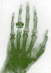 X-rays (or Röntgen rays) are a form of electromagnetic radiation with a wavelength in the range of 10 to 0.01 nanometers, corresponding to frequencies in the range 30 PHz to 30 EHz. X-rays are primarily used for diagnostic radiography and crystallography. X-rays are a form of ionizing radiation and as such can be dangerous. In many languages it is called Röntgen radiation after one of the first investigators of the X-rays, Wilhelm Conrad Röntgen. Units of Measure and ExposureThe rem is the traditional unit of Dose Equivalent. This describes the Energy delivered by γ or X - radiation (indirectly ionizing radiation) for humans. The SI counterpart is the Sievert (Sv). One Sievert is equal to 100 rem. Because the rem is a relatively large unit, typical equivalent Dose is measured in millirem (mrem), or one thousandth of a rem The average person living in the United States is exposed to approximately 150 mrem annually from background sources alone. Reported dosage due to dental X-rays seems to vary significantly. Depending on the source, a typical dental X-ray of a human results in an exposure of perhaps, 3, 40, 300, or as many as 900 mrems. Medical UsesSince Röntgen's discovery that X-rays can identify bony structures, X-rays have been developed for their use in medical imaging. Radiology is a specialized field of medicine. Radiographers employ radiography and other techniques for diagnostic imaging. Indeed, this is probably the most common use of X-ray technology. X-rays are especially useful in the detection of pathology of the skeletal system, but are also useful for detecting some disease processes in soft tissue. Some notable examples are the very common chest X-ray, which can be used to identify lung diseases such as pneumonia, lung cancer or pulmonary edema, and the abdominal X-ray, which can detect ileus (blockage of the intestine), free air (from visceral perforations) and free fluid (in ascites). In some cases, the use of X-rays is debatable, such as gallstones (which are rarely radiopaque) or kidney stones (which are often visible, but not always). Also, traditional plain X-rays pose very little use in the imaging of soft tissues such as the brain or muscle. Imaging alternatives for soft tissues are computed axial tomography (CAT or CT scanning), magnetic resonance imaging (MRI) or ultrasound. Since 2005, X-rays are listed as a carcinogen by the U.S. government. Radiotherapy, a curative medical intervention, now used almost exclusively for cancer, employs higher energies of radiation. The efficiency of X-ray tubes is less than 2%. Most of the energy is used to heat up the anode. Source: http://www.wikipedia.orgIf you find an error, please let us know.
Researched by Anthony Breast self-examination (BSE) is an easy but unreliable method for finding possible breast cancer. If performed appropriately and regularly BSE may help in early detection of some types of breast cancers, although it should not substitute for screening methods (such as mammography) that have been proven to be effective. The method involves feeling breasts for possible distortions or swelling. How to perform BSEThe steps involved in self exam are: · Stand in front of a mirror with top exposed. · Place hands on hips. · Look for signs of dimpling, swelling, soreness, or redness in all parts of your breasts in the mirror. · Repeat with arms raised above your head. · While still standing, palpate your breasts with your fingers, feeling for lumps. Try to use a larger area of your fingers rather than prodding. Feel both for the area just beneath the skin and for the tissue deeper within. · Go over the entire breast while examining. One method is to divide the breast into quadrants and palpate each quadrant carefully. Also examine the "axillary tail" of each breast that extends toward the axilla (armpit). · Repeat palpation while lying down. · Check the nipples and the area just beneath them. Gently squeeze each nipple to check for any discharge. See VideoThe Seven P's methodA similar method of self-examination is known as the Seven P's of BSE: 1. Position: Inspect breasts visually and palpate in the mirror with arms at various positions. Then perform the examination lying down, first with a pillow under one shoulder, then with a pillow under the other shoulder, and finally lying flat. 2. Perimeter: Examine the entire breast, including the nipple, the axillary tail that extends into the armpit, and nearby lymph nodes. 3. Palpation: Palpate with the pads of the fingers, without lifting the fingers as they move across the breast. 4. Pressure: First palpate with light pressure, then palpate with moderate pressure, and finally palpate with firm pressure. 5. Pattern: There are several examination patterns, and each woman should use the one which is most comfortable for her. The vertical strip pattern involves moving the fingers up and down over the breast. The pie-wedge pattern starts at the nipple and moves outward. The circular pattern involves moving the fingers in concentric circles from the nipple outward. Don't forget to palpate into the axilla. 6. Practice: Practice the breast self-exam and become familiar with the feel of the breast tissue, so you can recognize changes. A health care practitioner can provide feedback on your method. 7. Plan: Know what to do if you suspect a change in your breast tissue. Know your family history of breast cancer. Have mammography done as often as your health care provider recommends. For premenopausal women, BSE is best done at the same stage of their period every month to minimize changes due to the menstrual cycle. The recommended time is just after the end of the last period when the breasts are least likely to be swollen and tender. Older, menopausal women should do BSE once a month, perhaps on the first or last day of every month. About eight in ten lumps discovered by BSE are harmless. Nevertheless, any abnormality thus detected should immediately be reported to a doctor. Though most breast cancers are detected by women, BSE should be combined with an annual examination by a doctor for better chances of detection. Women can easily miss a breast lump that an expert can find. For the same reasons it is better to learn BSE from an expert. It is not a replacement for more trustworthy techniques like mammography or an examination using MRI. Note: consult a trustworthy site such as Johns Hopkins Breast Cancer Center for more complete and up-to-date information. Source: WikipediaIf you find an error, please let us know.
Researched by Anthony A laparoscopic tubal ligation can be done anytime when a woman is not pregnant. There are three ways to block the tube: Cautery (Burning); Applying Clips (Hulka Clips); Applying Silastic Rings. A LAPAROSCOPE is a wand shaped instrument used to see into the body. A small incision is made in the belly button (umbilicus) and gas is then infused into the abdomen. Once the abdomen is full of gas, the laparoscope is inserted. A second, puncture incision is made just above the pubic bone, while the surgeon is looking through the laparoscope (or watching on a television monitor if it is attached to a camera). A forceps-like instrument is then inserted into the abdomen through this incision. The forceps are used to grasp and elevate the tube. See Actual VideoThis is where the difference in the laparoscopic tubal comes in. CAUTERY: The ends of the forceps are two haves of an electrode and they squeeze the tube while an electric current is passed between them, burning the tube. This causes the most damage to the tube and is the least reversible of the three methods. The burn extends well beyond the area burned initially. CLIPS: The clip is held between the two sides of the forcep and the tube is squeezed between the halves, thus applying the clip. SILASTIC RINGS: The tube is grasped and pulled into the casing holding the forceps. This casing also holds a small ring a silastic material, much smaller than the tube. The tube is pulled through the ring and then dropped with the ring now in place and blocking the tube. This is done in an out patient surgery unit and you can go home in a few hours for rest. Postpartum Tubal Ligation Postpartum Tubal Ligations are done within two days of delivery because the top of the uterus is at the level of the belly button at that time. The Mom is put to sleep (If she has had an epidural for labor, the catheter may be left. She will then have epidural anesthesia). The belly button (umbilicus) is grasped and pulled up. A small incision is made in it. The tubes are pulled through this small incision one at a time using a grasping instrument that will not tear the tube. The tube is then tied securely with suture material. The tube is then cut, and usually the cut ends are cauterized. The tube is then replaced, gently, back into the abdomen. The same procedure is done for the second tube. After both tubes are cut, the belly button is sewn closed. Mom and baby can go home the next day. For c-section the tubes are tied and cut at the time of delivery. Misconceptions about Tubal LigationTubal Ligation does not affect the production and secretion of the female hormones, estrogen and progesterone. Therefore, there will be no masculinization developing such as voice deepening or hair growth; cessation of your period; early menopause; or decrease in libido (sex drive). Just because you had a tubal ligation does not mean you will have to have a hysterectomy some day. The days of unnecessary hysterectomy are dwindling. If a doctor advises one, always get a second opinion, REMEMBER: This is considered a PERMANENT method of birth control. While reversal is possible in some instances, it is not a guarantee. Source: http://www.mjbovo.comIf you find an error, please let us know.
 A petechia (IPA pronunciation: [pə'tiki.ə]), plural petechiae (IPA pronunciation: [pə'tiki.aɪ]) is a small red or purple spot on the body, caused by a minor hemorrhage (broken capillary blood vessels). Petechiae may be a sign of thrombocytopenia (low platelet counts). They also occur in circumstances when platelet function is inhibited (e.g., as a side effect of medications or during certain infections) or when excessive pressure is applied to tissue (e.g., when a tourniquet is applied to an extremity or with excessive coughing). Petechiae should always be quickly investigated. They can be interpreted as vasculitis, an inflammation of the blood vessels, which requires immediate treatment to prevent permanent damage. Some malignancies can also cause petechiae to appear. Petechiae should be investigated by a physician within a day or so to rule out the more dangerous conditions. Dermatologists can be the most helpful specialists in these conditions because they can more easily identify if the condition is petechiae or some similar looking but less worrisome rash. The significance of petechiae in children depends on the clinical context in which they arise. Petechiae in children can occur with viral infections. In this setting they do not necessarily signify serious illness. They are the hallmark of some possibly serious illnesses, however, such as meningococcemia, various causes of thrombocytopenia, and leukemia. Therefore, their presence should not be ignored. Associated Conditions * Bolivian hemorrhagic fever * Boutonneuse fever * Crimean-Congo hemorrhagic fever * Congenital syphilis * Dengue fever * Duke's disease * Ebola * Endocarditis * Erythroblastosis fetalis * Gua Sha * Henoch-Schönlein purpura * Leukemia * Childhood protein-energy malnutrition such as Kwashiorkor or Marasmus * Typhus * Scarlet Fever * Schamberg's Disease * Kawasaki disease * Kawasaki fever * Rocky mountain spotted fever Source: Wikipedia
Researched by Anthony A tourniquet test (also known as a Rumpel-Leede Capillary-Fragility Test or simply a capillary fragility test) determines capillary fragility. It is a clinical diagnostic method to determine a patient's hemorrhagic tendency. It assesses fragility of capillary walls and is used to identify thrombocytopenia (a reduced platelet count). The test is defined by the WHO as one of the necessary requisites for diagnosis of Dengue fever. A blood pressure cuff is applied and inflated to a point between the systolic and diastolic blood pressures for five minutes. The test is positive if there are more than 20 petechiae per square inch (a petechia is a small red or purple spot on the body, caused by a minor hemorrhage). This test does not have high specificity. Interfering factors with this test are women who are premenstrual, postmenstrual and not taking hormones, or those with sun damaged skin, since all will have increased capillary fragility.(Pagana, & Pagana, 1998; Tsai, 2000). ReliabilityAt least one insurance company, Aetna, has determined that the Rumpel-Leede test is obsolete or unreliable and has been replaced by more advanced procedures. (see http://www.aetna.com/cpb/data/CPBA0438.html.) The test remains an essential part of the assessment of a patient who may have dengue hemorrhagic fever. Source: Wikipedia - the Free Encyclopedia
Researched by Anthony In gynecology, the Papanikolaou test or Papanicolaou test (also called Pap smear, Pap test, cervical smear, or smear test) is a medical screening method, invented by Georgios Papanikolaou, primarily designed to detect premalignant and malignant processes in the ectocervix. It may also detect infections and abnormalities in the endocervix and endometrium. The endocervix may be partially sampled with the device used to obtain the ectocervical sample, but due to the anatomy of this area, consistent and reliable sampling cannot be guaranteed. As abnormal endocervical cells may be sampled, those examining them are taught to recognize them. The endometrium is not directly sampled with the device used to sample the ectocervix. Cells may exfoliate onto the cervix and be collected from there, so as with endocervical cells, abnormal cells can be recognized if present but the Pap Test should not be used as a screening tool for endometrial malignancy. The pre-cancerous changes (called dysplasias or cervical or endocervical intraepithelial neoplasia) are usually caused by sexually transmitted human papillomaviruses (HPVs). The test aims to detect and prevent the progression of HPV-induced cervical cancer and other abnormalities in the female genital tract by sampling cells from the outer opening of the cervix (Latin for "neck") of the uterus and the endocervix. The sampling technique changed very little since its invention by Georgios Papanikolaou (1883–1962) to detect cyclic hormonal changes in vaginal cells in the early 20th century until the development of liquid based cell thinlayer technology. The test remains an effective, widely used method for early detection of cervical cancer and pre-cancer. The UK's call and recall system is among the best; estimates of its effectiveness vary widely but it may prevent about 700 deaths per year in the UK. It is not a perfect test. "A nurse performing 200 tests each year would prevent a death once in 38 years. During this time she or he would care for over 152 women with abnormal results, over 79 women would be referred for investigation, over 53 would have abnormal biopsy results, and over 17 would have persisting abnormalities for more than two years. At least one woman during the 38 years would die from cervical cancer despite being screened." HPV vaccine may offer better prospects in the long term. It is generally recommended that sexually active females seek Pap smear testing annually, although guidelines may vary from country to country. If results are abnormal, and depending on the nature of the abnormality, the test may need to be repeated in three to twelve months. If the abnormality requires closer scrutiny, the patient may be referred for detailed inspection of the cervix by colposcopy. The patient may also be referred for HPV DNA testing, which can serve as an adjunct (or even as an alternative) to Pap testing. About 5% to 7% of pap smears produce abnormal results, such as dysplasia, possibly indicating a pre-cancerous condition. Although many low grade cervical dysplasias spontaneously regress without ever leading to cervical cancer, dysplasia can serve as an indication that increased vigilance is needed. Endocervical and endometrial abnormalities can also be detected, as can a number of infectious processes, including yeast and Trichomonas vaginalis. A small proportion of abnormalities are reported as of "uncertain significance". See Actual VideoTest PreparationsAn informed consent must be done prior to the procedure. The health care provider should be notified if the patient have had a prior abnormal Pap smear, if she might be pregnant, or if she's taking any medications or birth control pills. She should avoid douching, using tampons, having intercourse, and tub baths 24 hours before the test. Avoid scheduling Pap smear while the client is menstruating, because blood and cells from the endometrial cavity may obscure the accuracy of the Pap smear. Patient should empty her bladder just before the test. ConsiderationsThe following drugs may affect Pap smears: * Colchicine * Estrogen * Podophyllin * Progestins * Silver nitrate * Compounds in cigarettes Technical AspectsSamples are collected from the outer opening or os of the cervix using an Aylesbury spatula or (more frequently with the advent of liquid-based cytology) a plastic-fronded broom. The cells are placed on a glass slide and checked for abnormalities in the laboratory. The sample is stained using the Papanicolaou technique, in which tinctorial dyes and acids are selectively retained by cells. Unstained cells can not be visualized with light microscopy. The stains chosen by Papanicolau were selected to highlight cytoplasmic keratinization, which actually has almost nothing to do with the nuclear features used to make diagnoses now. The sample is then screened by a specially trained and qualified cytotechnologist using a light microscope. The terminology for who screens the sample varies according the country; in the UK, the personnel are known as Cytoscreeners, Biomedical scientists (BMS), Advanced Practitioners and Pathologists. The latter two take responsibility for reporting the abnormal sample which may require further investigation. Studies of the accuracy of conventional cytology report: · sensitivity 72% · specificity 94% In the United States, physicians who fail to diagnose cervical cancer from a pap smear have been convicted of negligent homicide. In 1988 and 1989, Karen Smith had received pap smears, which were argued to have "unequivocally" shown that she had cancer; yet the lab had not made the diagnosis. She died on March 8, 1995. Later, a physician and a laboratory technician were convicted of negligent homicide. These events have led to even more rigorous quality assurance programs, and to emphasizing that this is a screening, not a diagnostic test, associated with a small irreducible error rate. Liquid based monolayer cytology Since the mid-1990s, techniques based around placing the sample into a vial containing a liquid medium which preserves the cells have been increasingly used. The media are primarily ethanol based. Two of the types are Sure-Path (TriPath Imaging) and Thin-Prep (Cytyc Corp). Once placed into the vial, the sample is processed at the laboratory into a cell thin-layer, stained, and examined by light microscopy. The liquid sample has the advantage of being suitable for low and high risk HPV testing and reduced unsatisfactory specimens from 4.1% to 2.6%. Proper sample acquisition is crucial to the accuracy of the test; clearly, a cell that is not in the sample cannot be evaluated. Studies of the accuracy of liquid based monolayer cytology report: · sensitivity 61% to 66% · specificity 82% to 91% Some, but not all studies, report increased sensitivity from the liquid based smears. Human papillomavirus testing The presence of HPV indicates that the person has been infected, the majority of women who get infected will successfully clear the infection within 18 months. It is those who have an infection of prolonged duration with high risk types (e.g. types 16, 18, 31, 45) that are more likely to develop Cervical Intraepithelial Neoplasia due to the effects that HPV has on DNA. Studies of the accuracy of HPV testing report: · sensitivity 88% to 91% (for detecting CIN 3 or higher) to 97% (for detecting CIN2+) · specificity 73% to 79% (for detecting CIN 3 or higher) to 93% (for detecting CIN 3 or higher) By adding the more sensitive HPV Test, the specificity may decline. However, the drop in specificity is not definite. If the specificity does decline, this results in increased numbers of false positive tests and many women who did not have disease having colposcopy and treatment. A worthwhile screening test requires a balance between the sensitivity and specificity to ensure that those having a disease are correctly identified as having it and equally importantly those not identifying those without the disease as having it. Due to the liquid based pap smears having a false negative rate of 15-35%, the American College of Obstetricians and Gynecologists and American Society for Colposcopy and Cervical Pathology have recommended the use of HPV testing in addition to the pap smear in all women over the age of 30. Regarding the role of HPV testing, randomized controlled trials have compared HPV to colposcopy. HPV testing appears as sensitive as immediate colposcopy while reducing the number of colposcopies needed. Randomized controlled trial have suggested that HPV testing could follow abnormal cytology or could precede cervical cytology examination. A study published in April 2007 suggested the act of performing a Pap smear produces an inflammatory cytokine response, which may initiate immunologic clearance of HPV, therefore reducing the risk of cervical cancer. Women who had even a single Pap smear in their history had a lower incidence of cancer. "A statistically significant decline in the HPV positivity rate correlated with the lifetime number of Pap smears received." Automated analysis In the last decade there have been successful attempts to develop automated, computer image analysis systems for screening. Automation may improve sensitivity and reduce unsatisfactory specimens. One of these has been FDA approved and functions in high volume reference laboratories, with human oversight. Practical AspectsThe physician or operator collecting a sample for the test inserts a speculum into the patient's vagina, to obtain a cell sample from the cervix. A pap smear appointment is normally not scheduled during menstruation. The procedure is usually just slightly painful, because of the neuroanatomy of the cervix. However, this can depend on the patient's anatomy, the skill of the practitioner, psychological factors, and other conditions. Results usually take about 3 weeks. Slight bleeding, cramps, and other discomfort can occur afterwards. Other tests, including the TruTest, an endometrial biopsy used for early detection of uterine cancer, can be performed during the same visit. RisksThere are no risks involved. References:http://www.wikipedia.orghttp://www.nlm.nih.govIf you find an error, please let us know.
Researched by Anthony Thoracentesis Thoracentesis (also known as thoracocentesis or pleural tap) is an invasive procedure to remove fluid or air from the pleural space for diagnostic or therapeutic purposes. A cannula, or hollow needle, is carefully introduced into the thorax, generally after administration of local anesthesia. The procedure was first described in 1852. The recommended location varies depending upon the source. Some sources recommend the midaxillary line, in the sixth, seventh, or eighth intercostal space. See Actual VideoIndicationsThis procedure is indicated when unexplained fluid accumulates in the chest cavity outside the lung. In more than 90% of cases analysis of pleural fluid yields clinically useful information. If a large amount of fluid is present, then this procedure can also be used therapeutically to remove that fluid and improve patient comfort and lung function. The most common causes of pleural effusions are cancer, congestive heart failure, pneumonia, and recent surgery. In countries where tuberculosis is common, this is also a common cause of pleural effusions. When cardiopulmonary status is compromised (i.e. when the fluid or air has its repercussions on the function of heart and lungs), due to air (significant pneumothorax), fluid (pleural fluid) or blood (hemothorax) outside the lung, then this procedure is usually replaced with tube thoracostomy, the placement of a large tube in the pleural space. ContraindicationsAn uncooperative patient or a coagulation disorder that can not be corrected are absolute contraindications. Relative contraindications are site of insertion has known bullous disease (e.g. emphysema), use of positive end-expiratory pressure (PEEP) and only one functioning lung (due to diminished reserve). ComplicationsMajor complications are pneumothorax (3-30%), hemopneumothorax, hemorrhage, hypotension (low blood pressure due to a vasovagal response) and re-expansion pulmonary edema. Minor complications include a dry tap (no fluid return), subcutaneous hematoma or seroma, anxiety, dyspnea and cough (after removing large volume of fluid). Interpretation of pleural fluid analysisSeveral diagnostic tools are available to determine the etiology of pleural fluid. Transudate versus exudateFirst the fluid is either transudate or exudate. A transudate is defined as pleural fluid to serum total protein ratio of less than 0.5, pleural fluid to serum LDH ratio < style="font-style: italic;">Amylase A high amylase level (twice the serum level or the absolute value is greater than 160 Somogy units) in the pleural fluid is indicative of either acute or chronic pancreatitis, pancreatic pseudocyst that has dissected or ruptured into the pleural space, cancer or esophageal rupture. GlucoseThis is considered low if pleural fluid value is less than 50% of normal serum value. The differential diagnosis for this is: * rheumatoid effusion * lupus effusion * bacterial empyema * malignancy * tuberculosis * esophageal rupture (Boerhaave syndrome) pHNormal pleural fluid pH is approximately 7.60. A pleural fluid pH below 7.30 with normal arterial blood pH has the same differential diagnosis as low pleural fluid glucose. Triglyceride and cholesterolChylothorax (fluid from lymph vessels leaking into the pleural cavity) may be identified by determining triglyceride and cholesterol levels, which are relatively high in lymph. A triglyceride level over 110 mg/dl and the presence of chylomicrons indicate a chylous effusion. The appearance is generally milky but can be serous. The main cause for chylothorax is rupture of the thoracic duct, most frequently as a result of trauma or malignancy (such as lymphoma). Cell count and differentialThe number of white blood cells can give an indication of infection. The specific subtypes can also give clues as to the type on infection. The amount of red blood cells are an obvious sign of bleeding. Cultures and stainsIf the effusion is caused by infection, microbiological culture may yield the infectious organism responsible for the infection, sometimes before other cultures (e.g. blood cultures and sputum cultures) become positive. A Gram stain may give a rough indication of the causative organism. A Ziehl-Neelsen stain may identify tuberculosis or other mycobacterial diseases. CytologyCytology is an important tool in identifying effusions due to malignancy. The most common causes for pleural fluid are lung cancer, metastasis from elsewhere and mesothelioma. The latter often presents with an effusion. Normal cytology results do not reliably rule out malignancy, but make the diagnosis more unlikely. Source: Wikipedia - the Free EncyclopediaIf you find an error, please let us know.
h.s. or HS stands for hora somni, which means before sleep or at bedtime.
In pediatrics, the normal heart rates are: Infant: 100-160 bpm Toddler: 90-150 bpm Preschooler: 80-140 bpm School-age: 70-120 bpm If the heart rate goes above the normal range, this is already called Tachycardia. Source: http://www.health.state.ny.usIf you find an error, please let us know.
In pediatrics, the normal heart rates are: Infant: 100-160 bpm Toddler: 90-150 bpm Preschooler: 80-140 bpm School-age: 70-120 bpm If the heart rate goes below the normal range, this is already called Bradycardia. Note that a drop in heart rate may still be considered normal if the heart rate returns to normal within five to 10 seconds without any aid. Source: http://www.health.state.ny.usIf you find an error, please let us know.
Researched by Anthony A normal adult heart beats between 60 and 100 times a minute. A heart rate over 100 beats a minute is called tachycardia. Some tachycardias are relatively harmless and need no treatment, but others can be life-threatening. Treatment for recurrent tachycardia can range from daily medication to open-heart surgery. A specific diagnosis is necessary before finding the right treatment for the tachycardia. Signs and symptomsWhen the heart rate is too rapid, it may not effectively pump blood to the rest of the body, depriving organs and tissues of oxygen. This can cause these signs and symptoms: · Dizziness · Shortness of breath · Lightheadedness · Rapid heartbeat · Heart palpitations — a racing, uncomfortable or irregular heartbeat or a sensation of "flopping" in the chest · Chest pain · Blackouts · Visual problems · Fainting (syncope) Some people with tachycardia have no symptoms and don't realize they have this condition until a doctor discovers it during a physical examination. CausesThe heart is a muscular pump that circulates blood all around the body. There are four hollow chambers in the heart — the two upper chambers are the atria, and the lower, more muscular chambers are the ventricles. Each heartbeat begins in the right atrium. There, the heart's natural pacemaker, called the sinus node, sends an electrical signal that causes the atria to contract, filling the ventricles with blood. A split second later, the electrical impulse travels across the atrioventricular (AV) node into the ventricles. This makes the ventricles contract, sending blood throughout the body. In people with tachycardias, this normal rhythm is disrupted somewhere along the electrical path, causing the heart to beat too quickly. Types of TachycardiasTachycardias are classified according to where they originate — in the atria or in the ventricles. Tachycardias originating in the upper heart chambers Atrial fibrillation. In this most common arrhythmia in the U.S., electrical impulses make the atria beat extremely quickly — up to 400 beats a minute. Only some of these electrical impulses travel across the AV node and reach the ventricles, causing a rapid and irregular heartbeat. This tachycardia is most common in people over 60 years of age. Atrial flutter. Atrial flutter is similar to atrial fibrillation, except the extremely fast beating is more controlled and rhythmic. The most common symptom of atrial flutter is chest pain. Supraventricular tachycardia (SVT). SVT is a broad term that includes many forms of arrhythmia originating above the ventricles (supraventricular). SVTs usually cause a burst of rapid heartbeats that begin and end suddenly and can last from seconds to hours. These often start when the electrical impulse from a premature heartbeat begins to circle repeatedly through an extra pathway. SVT may cause the heart to beat 160 to 200 times a minute. Although generally not life-threatening in an otherwise normal heart, symptoms from the racing heart may feel quite uncomfortable. These arrhythmias are common in young people. Tachycardias originating in the ventricles Because the ventricles supply blood to the entire body, a tachycardia that starts in the ventricles can be a medical emergency. Types include: Ventricular tachycardia. This is a rapid, rhythmic heartbeat that most often affects people with structural heart disease with damage to the heart muscle (myocardium), such as occurs with a heart attack. Ventricular tachycardia can be life-threatening by itself, and without treatment it can rapidly turn into fatal ventricular fibrillation. Ventricular fibrillation. During ventricular fibrillation, rapid, chaotic electrical impulses cause the ventricles to quiver uselessly instead of pumping necessary blood to the body. This serious malfunction results in death if the heart isn't restored to a normal rhythm within minutes. Tachycardia TriggersIn some people, external substances can affect the heart's electrical system and cause a tachycardia to develop. People with sensitivities to the substances can develop tachycardias after moderate exposure, but abuse of these substances can also cause the arrhythmia directly. Substances include: * Caffeine * Alcohol * Tobacco * Dietary supplements and over-the-counter medications * Illicit drugs * Prescription drugs * Environmental pollutants, such as automobile emissions and paint thinner Risk factorsCertain factors can increase the risk of developing tachycardias. They include: Coronary artery disease. Hardening or narrowing of the heart's arteries, a previous heart attack or heart damage puts one at higher risk of developing an arrhythmia. Damaged heart muscle (cardiomyopathy). When cardiomyopathy damages the heart muscle, the electrical pathways can be affected. Damaged heart valves. The heart valves can become damaged due to cardiovascular disease, increasing the tachycardia risk. Older age. Aging-related wear on the heart makes one more susceptible to developing an arrhythmia. Genetics. If a person have a family history of arrhythmia disorders or heart disease, that person is at higher risk. Overactive thyroid (hyperthyroidism). An overactive thyroid gland releases excess hormones, causing metabolism to speed up. This can lead to fast or irregular heartbeats. Sleep apnea. When this sleep disorder causes one to stop breathing repeatedly during sleep, the lack of oxygen can lead to bursts of atrial fibrillation. Electrolyte imbalance. An imbalance of minerals in the blood, such as potassium, sodium, calcium and magnesium can affect the heart's electrical system, leading to irregular heartbeats. High blood pressure (hypertension). High blood pressure, especially if poorly controlled, puts a strain on the heart and can result in enlargement of the heart chambers or weakness of the heart muscle with an increased risk of tachycardia or both. Screening and DiagnosisTo diagnose and treat an arrhythmia, the doctor needs to document the type of abnormal rhythm. This can be done by monitoring the heart of the patient in the doctor's office or hospital or as he/she go about his/her daily activities, or by actively triggering tachycardia while being closely watched by a doctor. Tests that monitors heart rate Electrocardiogram (ECG). Sensor pads with wires attached, called electrodes, are placed on the chest and sometimes, the limbs to measure the heart's electrical impulses. Holter monitor. This small, portable ECG device is worn for 24 hours of continuous monitoring of the heart's electrical signals. Event monitor. An event monitor can help doctors evaluate more sporadic tachycardias. A patient wears this portable ECG device at home and activate it when he's/she's experiencing symptoms. The doctor can then evaluate the ECG strip to determine if there's an association between the symptoms and the heart's rhythm. Tests that trigger the arrhythmia include Stress test. Some tachycardias are triggered or made worse with exercise. During a stress test, a patient is made to walk or run on a treadmill, or ride a stationary bike, while his/her heart's rhythm is monitored with an electrocardiogram. Stress tests also try to determine if there is coronary artery disease. Electrophysiological mapping and testing. While a patient is under light sedation, a specialist in heart rhythm disorders (electrophysiologist) threads thin, flexible tubes (catheters) tipped with electrodes through the patient's blood vessels to various places inside the heart. From there, the electrodes precisely map the heart's electrical impulses. During testing, the electrodes stimulate the heart to trigger or halt the tachycardia, revealing its type and location. Tilt table test. This test may be used if a patient have recurrent fainting spells. A patient is made to lie flat on a special table, and then the table is tilted as if he/she is standing up. The doctor observes how the heart and nervous system respond to these changes in position. Tests that check the structure and function of the heart Echocardiogram. This test uses soundwaves to produce detailed images of the heart's structure, size and motion, which can give the doctor valuable information about arrhythmia. Coronary angiogram. During coronary angiography, a type of dye that's visible by X-ray machine is injected into the blood vessels of the heart. The X-ray machine rapidly takes a series of images (angiograms), offering a detailed look at the inside of blood vessels to see if coronary artery disease is causing a tachycardia. ComplicationsIn a heart arrhythmia, heartbeat irregularities may put one at higher risk of developing blood clots in the heart, which can dislodge and lead to a heart attack or stroke. Some people with known arrhythmias take blood-thinning medications called anticoagulants to prevent blood clots from forming. Tachycardias can be mild and cause no symptoms. They can also happen suddenly with potentially serious and even fatal results. Even a relatively harmless tachycardia can quickly turn into something deadly, or at least predict that one would have a more significant arrhythmia in the future. Proper diagnosis and treatment are key in preventing these complications. TreatmentTreatment of a tachycardia depends on the symptoms, how often a person have episodes of tachycardia and whether they have any underlying heart conditions. The doctor may prescribe medications to control the heart rate, use minimally invasive procedures to destroy tachycardia trigger spots, or suggest the placement of a heart device that monitors and resets the heart rhythm as needed. Medications
Doctors often use medications in tachycardia emergencies, and they also prescribe them for long-term control of the arrhythmia. Medications may be used alone or in combination with other treatments. Anti-arrhythmic medications. These medications work to slow the conduction of electrical signals or prolong the time it takes the heart muscle to recharge after a beat. In some people with tachycardia, however, these medications can make arrhythmias worse. Calcium channel blockers, potassium channel blockers and beta blockers. These medications work in various ways to slow nerve impulses in the heart muscle or reduce the workload on the heart. These medications are also used to treat high blood pressure, coronary artery disease and chest pain. Blood thinners. Also called anticoagulants, these medications are not used to treat the abnormal heart rhythm, but rather to reduce the risk that the abnormal rhythm will trigger a blood clot that may lead to a stroke or heart attack. Anticoagulants make it more difficult for blood to clot. Nonsurgical Procedures Nonsurgical techniques may be used if medications don't work. Some examples include: Cardioversion. Most effective for atrial tachycardias, this technique uses either drugs or an electrical shock to restore a normal rhythm. In electric cardioversion, the doctor shocks the heart to stop its beat for a split second to "reset" it. Cardioversion drugs may be given through an intravenous (IV) line to reset the heart. Radiofrequency catheter ablation. In this procedure done under mild sedation, several electrode-tipped tubes (catheters) are threaded through the blood vessels to specific electrical pathways in the heart. Then radiofrequency energy is used to destroy the area of heart tissue responsible for the arrhythmia. Implantable cardioverter-defibrillator If the underlying heart condition puts one at high risk of having a life-threatening tachycardia, the doctor may recommend an implantable cardioverter-defibrillator (ICD). An implantable cardioverter-defibrillator — a pager-sized device implanted in the chest like a pacemaker — detects and stops dangerous arrhythmias. The ICD continuously monitors the heartbeat and delivers precisely calibrated electrical shocks to restore a normal heart rhythm. Surgical treatments If a patient does not respond to other treatments or needs a treatment for an underlying heart condition, surgery may be an option. Maze procedure. The maze procedure is often done during an open-heart surgery. Using a scalpel, doctors create several precise incisions in the atria to create a pattern or maze of scar tissue. Because scar tissue doesn't carry electricity, it interferes with stray electrical impulses that cause atrial fibrillation. Radiofrequency or cryotherapy also can be used, and there are several variations of the surgical maze technique. The procedure has a high success rate, but because it usually requires open-heart surgery, it's generally reserved for people who don't respond to other treatments or when it can be done during other necessary heart surgery, such as coronary artery bypass surgery or heart valve repair. Some people require a pacemaker after the procedure. PreventionThe most effective way to prevent tachycardias is to reduce the risk of developing heart disease. If one already have heart disease, he/she needs to monitor it and follow his/her treatment plan to lower his/her tachycardia risk. Prevent heart disease Treat or eliminate risk factors that may lead to heart disease. The following steps may be taken: Exercising and eating a healthy diet. Living a heart-healthy lifestyle by exercising regularly and eating a healthy, low-fat diet that's rich in fruits, vegetables, whole grains and antioxidants. Maintaining a healthy weight. Obesity puts one at higher risk of developing heart disease and tachycardias. Keeping blood pressure and cholesterol under control. Making lifestyle changes or taking medications to correct high blood pressure (hypertension) or high cholesterol. Cessation of smoking. Tobacco use increases the risk of cardiovascular disease and heart arrhythmias. Stress Control. Avoiding unnecessary stress, and learning coping techniques to handle normal stress in a healthy way. Constant Checkups. Having regular physical exams and reporting any signs or symptoms to a doctor. Monitor and treat existing heart disease If a person already have heart disease, there are steps he/she can take to lower his/her risk of developing a tachycardia or other arrhythmia: Following the plan. Understanding the treatment plan, and taking all medications as prescribed. Getting electrical system checked. One should talk to a doctor about whether a heart attack or another cause has compromised the heart's electrical system and put him/her at risk of sudden cardiac death. Reporting changes immediately. If symptoms change, get worse or develop new symptoms, one should tell his/her doctor immediately. Coping skillsIf a person have tachycardia, he/she may feel like his/her heart could spin out of control any second. This uncertainty may leave him/her feeling anxious and stressed. The best thing he/she can do is to follow his/her treatment plan and talk with his/her doctor about any concerns he/she may have. It may also help to meet with a psychologist, a genetics counselor or a cardiologist. Source: http://www.mayoclinic.comIf you find an error, please let us know.
Researched by Anthony Bradycardia Bradycardia, as applied to adult medicine, is defined as a resting heart rate of under 60 beats per minute, though it is seldom symptomatic until the rate drops below 50 beat/min. Trained athletes tend to have slow resting heart rates, and resting bradycardia in athletes should not be considered abnormal if the individual has no symptoms associated with it. The term relative bradycardia is used to explain a heart rate that, while not technically below 60 beats per minute, is considered too slow for the individual's current medical condition. This cardiac arrhythmia can be underlain by several causes, which are best divided into cardiac and non-cardiac causes. Non-cardiac causes are usually secondary, and can involve drug use or abuse; metabolic or endocrine issues, especially in the thyroid; an electrolyte imbalance; neurologic factors; autonomic reflexes; situational factors such as prolonged bed rest; and autoimmunity. Cardiac causes include acute or chronic ischemic heart disease, vascular heart disease, valvular heart disease, or degenerative primary electrical disease. Ultimately, the causes act by three mechanisms: depressed automaticity of the heart, conduction block, or escape pacemakers and rhythms. CausesThere are generally two types of problems that result in bradycardias: disorders of the sinus node, and disorders of the atrioventricular node (AV node). With sinus node dysfunction (sometimes called sick sinus syndrome), there may be disordered automaticity or impaired conduction of the impulse from the sinus node into the surrounding atrial tissue (an "exit block"). It is difficult and sometimes impossible to assign a mechanism to any particular bradycardia, but the underlying mechanism is not clinically relevant to treatment, which is the same in both cases of sick sinus syndrome: a permanent pacemaker. Atrioventricular conduction disturbances (aka: AV block; 1° AV block, 2° type I AV block, 2° type II AV block, 3° AV block) may result from impaired conduction in the AV node, or anywhere below it, such as in the bundle of HIS. Patients with bradycardia have likely acquired it, as opposed to having it congenitally. Bradycardia is more common in older patients. ManagementThere are two main reasons for treating any cardiac arrhythmias. With bradycardia, the first is to address the associated symptoms, such as fatigue, limitations on how much an individual can physically exert, fainting (syncope), dizziness or lightheadedness, or other vague and non-specific symptoms. The other reason to treat bradycardia is if the person's ultimate outcome (prognosis) will be changed or impacted by the bradycardia. Treatment in this vein depends on whether any symptoms are present, and what the underlying cause is. Primary or idiopathic bradycardia is treated symptomatically if it is significant, and the underlying cause is treated if the bradycardia is secondary. TreatmentDrug treatment for bradycardia is typically not indicated for patients who are asymptomatic. In symptomatic patients, underlying electrolyte or acid-base disorders or hypoxia should be corrected first. IV atropine may provide temporary improvement in symptomatic patients, although its use should be balanced by an appreciation of the increase in myocardial oxygen demand this agent causes. Source: Wikipedia - the Free EncyclopediaIf you find an error, please inform us.
|
 X-rays (or Röntgen rays) are a form of electromagnetic radiation with a wavelength in the range of 10 to 0.01 nanometers, corresponding to frequencies in the range 30 PHz to 30 EHz. X-rays are primarily used for diagnostic radiography and crystallography. X-rays are a form of ionizing radiation and as such can be dangerous. In many languages it is called Röntgen radiation after one of the first investigators of the X-rays, Wilhelm Conrad Röntgen.


